Researchers get to the heart of organ development
18 May 2025

Advanced microscopy has enabled researchers to capture in real time the formation of an animal heart.
Scientists from UCL and the Francis Crick Institute used 3D imaging to reveal the two day process by which individual cells in a mouse embryo began to form the vital organ.
Senior author Dr Kenzo Ivanovitch, of UCL Great Ormond Street Institute of Child Health and intermediate research fellow at the British Heart Foundation, explained the unique event.
“This is the first time we’ve been able to watch heart cells this closely, for this long, during mammalian development. We first had to reliably grow the embryos in a dish over long periods, from a few hours to a few days, and what we found was totally unexpected.”
The researchers outlined in The EMBO Journal how they employed advanced light-sheet microscopy on an engineered mouse model.
They combined this technique with tagging cardiomyocyte heart muscle cells with fluorescent markers, taking images every two minutes for more than 40 hours.
By tracking them back to earlier cells, the scientists identified when the first cells engaged only in forming the heart appeared within the embryo.
Focussing on the period of gastrulation – when cells specialise and organise into the body’s primary structures – they discovered that four to five hours after the first cell division, cells contributing solely to the heart emerged and followed distinct rather than random patterns.
Ivanovitch said: “This fundamentally changes our understanding of cardiac development by showing that what appears to be chaotic cell migration is actually governed by hidden patterns that ensure proper heart formation.”
The research offers potential aid in the study of congenital heart defects in humans – conditions that affect an estimated one in a hundred newborn babies, say the scientists.
Lead author, PhD candidate Shayma Abukar of UCL Great Ormond Street Institute of Child Health and UCL Institute for Cardiovascular Science said: “We are now working to understand the signals that coordinate this complex choreography of cell movements during early heart development.”
It is also hoped that the work could assist the development of lab grown heart tissue techniques necessary for regenerative medicine.
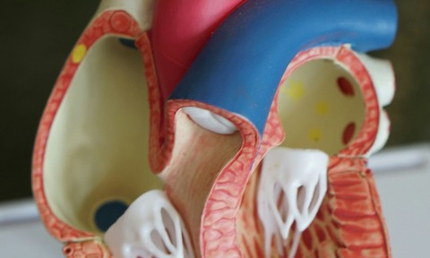
Studies of animal heart development could pave the way for improved understanding of the human heart (Pic: Robina Weermeijer)

